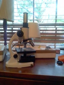
Model and name of the instrument: Student school biological microscope. RM-1B Radical Instruments
What is it used for? Microscopes are used for viewing very small objects, like insects or cells, up close.
Who needs it? Microscopes can be used by many different people, but this one is made specifically for students.
What parts is it made of? The microscope has an eyepiece, a body tube, and arm, a stage, 3 objective lenses, and a revolving nose piece to switch the objective lens. Also, it has stage clips to clip the viewing slides to the stage, some focus adjustment knobs, a base, and a mirror for illumination.
How does it work? Describe in great detail. You look through the eyepiece and you can see the slide through one of the objective lenses. The mirror or light under the stage illuminates the slide. You can focus the image by turning the focus adjustment knobs on the sides of the microscope.
Read the details on what is Histology, how H&E works, how to identify different type of cells and tissue (how different disease cells/tissues look?):
A Beginner’s Guide to Haematoxylin and Eosin Staining
https://histology.leeds.ac.uk/what-is-histology/H_and_E.php
Katya:
This is the microscope. It is working. There is nothing missing.
June 29, 2020
HISTOLOGY: Histology, which can also be known as microanatomy, is the microscopic study of cells and tissues. Histopathology is a branch of histology that studies diseased tissues in medicine.
H&E: Tissues and cells are transparent of dull grey in nature, which makes them really hard to view under a microscope. For this reason, the cells and tissues are stained with different colors. The most common staining system is H&E, which consists of the dyes Hematoxylin and Eosin.
Eosin is an acidic dye which is negatively charged. Because acidic dyes bind to basic cell structures, Eosin dyes cytoplasm, which is basic, a red or pink color.
Hematoxylin is a basic dye, so it stains acidic structures purple or blue. Hematoxylin is actually a dye called hematein. Hematein alone cannot stain tissues. It needs to be used together with a mordant, which is a compound. When hematein stains something, the mordant first binds to the tissue, then the hematein binds to the mordant.
June 30, 2020
IDENTIFYING DISEASED TISSUE: Histology can be helpful when a doctor is trying to diagnose a certain disease. Some diseases have clear visual signs of their presence, while others require non-histological forms of testing.
Even though we usually can’t see the virus or bacteria itself under a light microscope, we can still see the signs that the virus is present. The tissue around the area will be damaged, and certain spots could be colored differently under H&E staining. That is a spot impacted by a virus or bacteria.
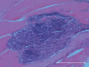
This is not my photo, see Introduction to histopathology.
July 1, 2020
Dima, you told me to look at Maria’s work on a similar project to help me figure out what to do next. I will continue to do the following:
- Research morphology of cells (how to identify different kinds of cells)
- Research architecture of tissues (how cells are structured in tissues)
- learn how the structure of cells and tissues affects their function
- Learn how cells and tissues appear normally
- learn how cells and tissues change during disease
- identify which slides have diseased cells and which have healthy cells
MORPHOLOGY: the branch of biology that deals with the form of living organisms, and with relationships between their structures. (definition from Google)
July 2, 2020
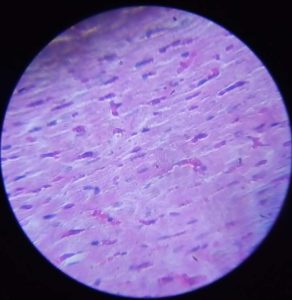
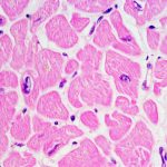
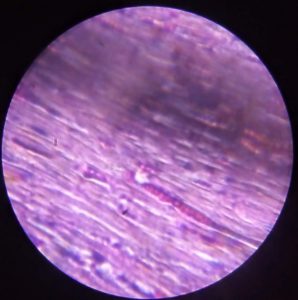
This is a picture I took. These are skeletal muscle cells. The little purple dots are nuclei.
July 3rd and 4th, 2020
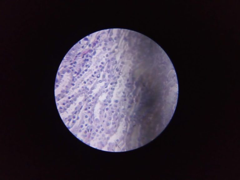
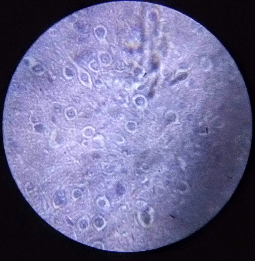
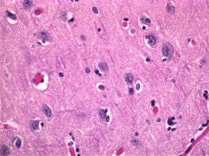
I think these maybe brain cells.
July 5, 2020
Dima, I read your instructions and will now be posting examples of each type of cell here, before I try to find them on my slides.
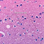
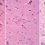
BRAIN CELL EXAMPLES

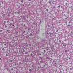
LIVER CELL EXAMPLES
LUNG TISSUE EXAMPLES
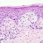
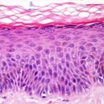
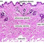
SKIN TISSUE EXAMPLES
KIDNEY TISSUE EXAMPLES
HEART TISSUE EXAMPLES
July 9, 2020
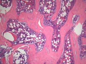
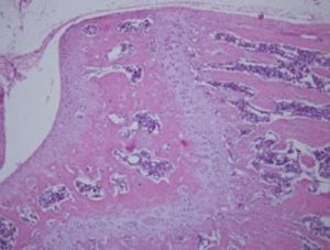
BONE TISSUE EXAMPLES
July 10, 2020
Dima, I have put examples of a lot of different tissues here already. I have also taken photos of most of my slides, so I will now try to identify those photos. When I am left with only ones that I don’t know, I will continue posting examples. Some of my photos turned out with really bad quality because my microscope isn’t ideal for taking photos through it, so I will be retaking some. Here are the ones that turned out ok, and I know what they might be.
MY PHOTOS
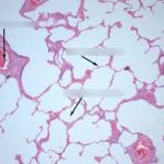
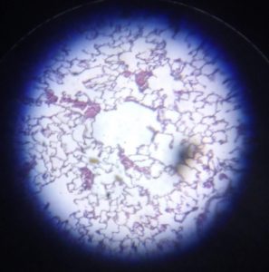
I think this is lung tissue
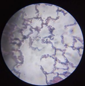
same as above

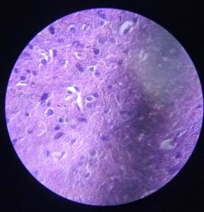
I think these could be kidney cells, though I am not sure due to the fact that photo quality isn’t very good. It looks much clearer just looking through the microscope.
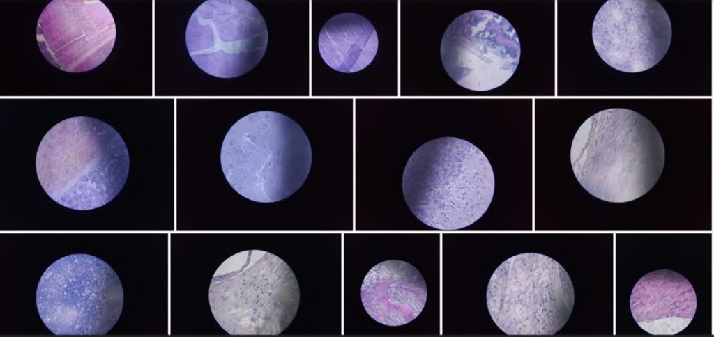
I am going to retake some images because they are very blurry. Also, I will need to post more examples. Here are some photos I have that I am not sure of:
July 13, 2020
I wanted to post more examples because I am not sure if these are the only ones in the slides. But I couldn’t find good pictures for a couple of different tissues. So for now I will just retake some of the pictures that didn’t turn out very well.
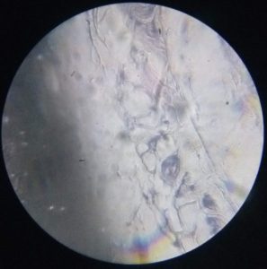
Also, a lot of slides have something like this on it. Could you maybe help me figure out what it is?
July 14, 2020
Dima, you told me that I wasn’t doing exactly what you wanted me to do, so from now on, I will begin to post examples and identify cells one by one.
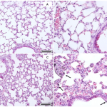
LUNG CELLS:
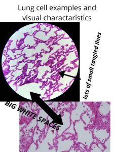
The little purple dots that are hard to see are nuclei. The things that I labeled here are simply visual characteristics that help identify the tissue.
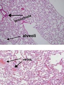
I think that lung cells have a very unique shape and structure, so they are fairly easy to identify.
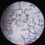
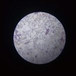
lung cells found in my slides:
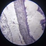
This is also lung tissue because of the bit that’s on the left side. The part in the middle is probably some kind of muscle.
I have a question: While the shape and structure of the example lung cells and the lung cells from my slides are the same, the coloring is different. Does that mean that they were stained differently? Or is it just my microscope? Or maybe they are in different conditions?
July 14, 2020
BRAIN CELLS:
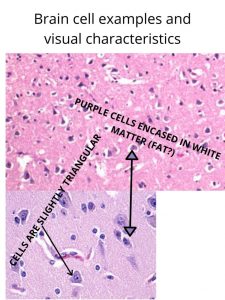
All of these photos are not mine. the labels on the larger photo above these two are not mine either.
July 15, 2020
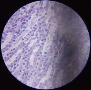
Brain cells found in my slides:
A lot of these look a little bit different but I think these are brain cells.
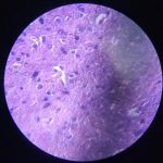
I’m sorry about the shadow on the sides of the photos. I think this has something to do with the microscope’s objective lens, which I have tried to clean, but I can’t fix it.

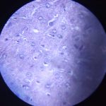
These a look a little bit different than the ones above, so I am not 100% sure that they are the same type of cells.
July 16, 2020
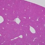
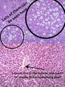
Liver Cells:
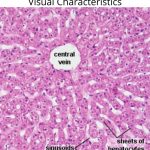
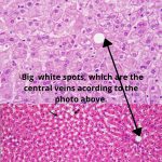
Actually, (I realized this after I made the labeled photos) in the top photo here, the white spot is NOT a central vein, because that photo is taken with higher magnification than the very first photo. The white spot is probably a smaller vein or one of the sinusoids labeled in the first picture.
July 21, 2020
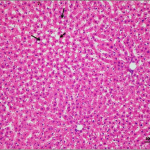
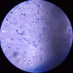
liver cells found in my slides:
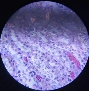
This is pretty blurry, but I think this is about the same magnification as the very first picture. This is the only one that I could find so far. I will keep looking.
July 23, 2020
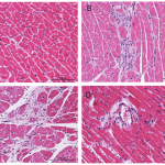
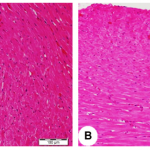
MUSCLE CELLS:
There are 3 kinds of muscle cells.
- Skeletal Muscles. These are the muscles on your bones that work when you consciously move, like when walking.
- Smooth Muscles. These muscles help organs like your stomach work.
- Cardiac Muscles. These muscles are what makes your heart pump blood.
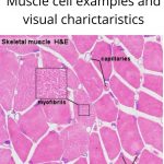
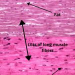
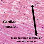
These are not my labels in the photo above.
7/26/2020: From Dima: Internship deadline is August 17, 2020. You have 3 weeks left, please note that it will not be possible to add text/data after this date due to the nature of summer internship and associated time limits.
July 28, 2020
Muscle cells found in my slides:

This is either skeletal or smooth muscle.
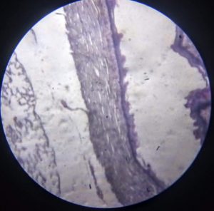
This is slide has some lung tissue on the left, but I am pretty sure the tissue in the middle is muscle. Since it’s muscle in an organ, it is probably smooth muscle. I hope this is correct.
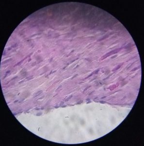
This is either skeletal or smooth muscle.
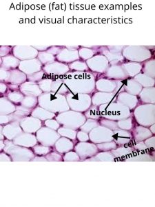
ADIPOSE TISSUE:
Adipose tissue is fat, a connective tissue that insulates and cushions the body.
It is pretty easy to identify, due to the fact that it looks nothing like most other tissue.
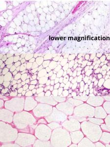
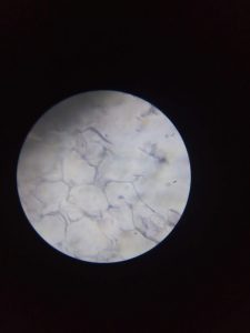
Adipose cells found in my slides:
Same slide with lower magnification.
August 6, 2020
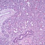
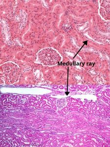
KIDNEY TISSUE:
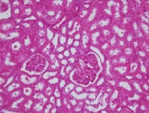
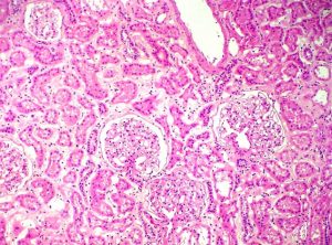
Kidney tissue examples:

The Glomerulus, plural Glumeri, is a group of capillaries (tiny blood vessels) where waste is filtered from the blood.
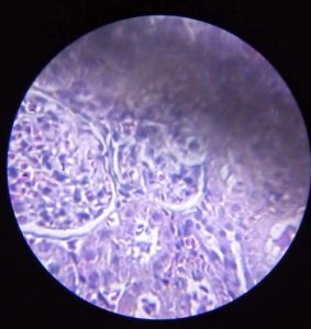
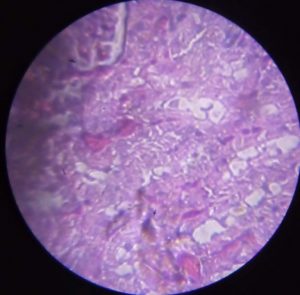
kidney cells found in my slides:
August 12, 2020
The four basic types of tissue:
I already knew some of this but I decided to add this information to better learn and understand it.
- Epithelial tissue: This tissue is what makes up skin and lines the inside of various organs
- connective tissue: This tissue connects and supports other tissue. Bone, blood, adipose(fat), and lymph tissue are all connective
- note: lymph or lymphoid tissue is part of the lymphatic system. Its main function is to transport white blood cells through the body. Similar to the immune system.
- Muscle tissue: Each and every little muscle in your body
- Nerve tissue: Made of nerve cells and is used to carry messages throughout the body.
Information on this found here.
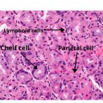
STOMACH TISSUE:
Also know as parietal cells, stomach cells are epithelium cells that can produce hydrochloric acid (stomach acid).
Gastric chief cells produce pepsinogen and chymosin. Pepsinogen largely helps digest proteins, while chymosin curdles and digests milk products.
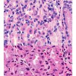
Stomach tissue examples:
August 13, 2020
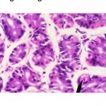
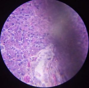
stomach tissue found in my slides:
References, citations, links:
I couldn’t post all the links because some of them didn’t work. For all the example photos I just typed into Google the tissue type and then H&E or histology.
https://www.webpathology.com/image.asp?case=678&n=3
https://en.wikipedia.org/wiki/Parietal_cell
https://en.wikipedia.org/wiki/Parietal_cell
https://librepathology.org/wiki/Proton_pump_inhibitor_effect
http://histology-world.com/photoalbum/displayimage.php?album=15&pid=714
https://renaissance.stonybrookmedicine.edu/pathology/neuropathology/chapter1
https://ntp.niehs.nih.gov/nnl/nervous/brain/microgliosis/brain-microgliosis_508.pdf
https://www.kidneypathology.com/English_version/Case_84.html