Model and name of the instrument: Microscope, Olympus BHTU / BH-2
What is it used for? To view samples on slides and see observe the samples to see things that would be otherwise impossible to see with the naked eye.
Who needs it? Scientists and manufacturers, because scientists can look at samples up close to learn more about the said samples and make observations, and manufacturers need them to inspect their products before selling them to find faults.
What parts is it made of? It is made from mostly lenses, gears metal, plastic, and electronic parts for the light.
How does it work? Describe in great detail. Using the lenses inside the microscope to refract the light that passes through the slides that are being viewed so that when you look into the microscope, you see the magnified image, which allows you to then make observations. There are special handles connected to gears within the microscope that enable you to make large or small adjustments to the distance between the lense and slide that you are observing, which helps to make the magnification of the slide clearer or blurrier. You can switch between lenses and doing so will allow you to observe at a higher or lower magnification.
Were you able to find an instrument manual online? I was not able to, however, I already have a lot of experience with microscopes since childhood.
Read the manual. Describe what was clear in the manual and what was confusing. I in fact, also have a microscope and slides of a large diversity, so I can make even more observations.
I was able to get the microscope to work, and was able to adjust between lenses and distance between lense and slide perfectly fine, and the light was also working just fine. I took these pictures myself by putting my phone to the lenses
Very healthy and vibrant looking cells, I have yet to find out what this is.
6/28/20 Edit: there seems to be a lot of epithelium and adipose, I don’t yet know what this is but it probably is the lining of a hollow organ like blood vessels or intestines. I added labels to the picture:
Tadaaa!
All is well with the microscope.
This is from the samples on the left side of the slide, and it is clearly the opposite of the samples on the right, and the cells seem damaged and are not as vibrant as the others.
Some more of the same sample as the previous picture, which were taken from the samples to the right of the slide.
Left: damaged
Right: healthy
I am going to research histology on the internet. Luckily, I found multiple websites that explain histology and the identification of tissues and what you see under the microscope.
http://www.histologyguide.com/slidebox/slidebox.html
https://www.researchgate.net/publication/283490690_Junqueira’s_Basic_Histology_Text_Atlas_14th_ed
I fixed the pictures! – Maria
Using this excerpt of the internship description, I will determine my learning goals:
Histology is the study of the structures of cells and tissues. Student will learn about the morphology of cells and the architecture of tissues, as well as how those structures indicate and facilitate the functions of cells and tissues. Histology focuses on the normal, how cells and tissues should appear in the absence of disease or infection. Understanding the normal will allow student to identify changes that occur to cells and tissues during disease.
Student will learn basics of the most commonly used staining system that is called H&E (Haemotoxylin and Eosin). Student will get microscope and hundreds of slides for identification of cells (muscle and brain cells, fibroblasts, astrocytes and adipocytes), cell compartments (nucleus, cytoplasm, ribosomes, endoplasmic reticulum, intracellular membranes), extracellular fibres and materials (carbohydrates in cartilage), and various types of H&E stained tissues. Eosin is an acidic dye: it is negatively charged and stains basic (or acidophilic) structures red or pink (e.g. cytoplasm). Haematoxylin is used to stain acidic (or basophilic) structures a purple-blue.
Learning goals:
- morphology of cells
- architecture of tissues
- how those structures indicate and facilitate the functions of cells and tissues
- how cells and tissues should appear in the absence of disease or infection, normal
- identify changes that occur to cells and tissues during disease
- learn basics of the most commonly used staining system that is called H&E (Haemotoxylin and Eosin)
- identify what slide is Haemotoxylin stained and what slide is Eosin stained
What is included in the slides: cells (muscle and brain cells, fibroblasts, astrocytes and adipocytes), cell compartments (nucleus, cytoplasm, ribosomes, endoplasmic reticulum, intracellular membranes), extracellular fibers and materials (carbohydrates in cartilage), and various types of H&E stained tissues (Just to know what to consider when trying to identify slide)
Notes from research:
Adipocytes – fat cells
If there is a big white spot where there is epithelium closed around it, it may be a blood vessel. For example, the below photos depict adipose tissue:
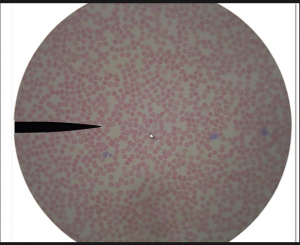
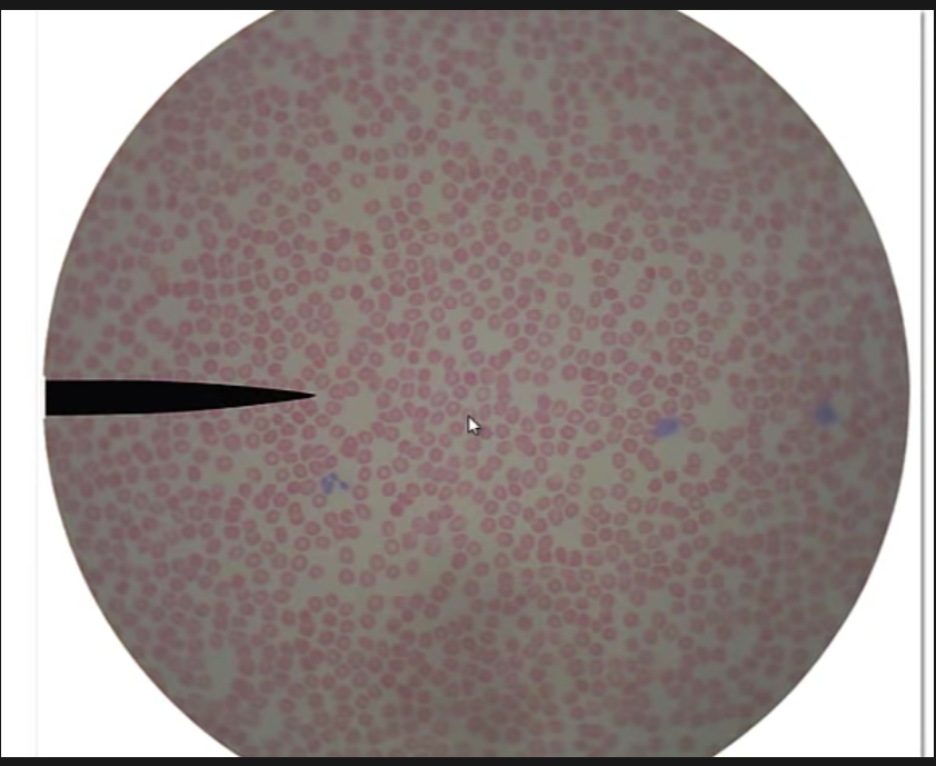
This is blood, the dots are red blood cells/ erythrocytes. The bluish stained dots are white blood cells, the white blood cells usually stain blue/ purple. There are also platelets which play a role in stopping bleeding or blood clotting. The platelets are darker colored than the red blood cells, but are still hard to spot.
Note: look up blood histology.
Hemostasis – the stopping of the flow of blood, notice the word stasis in there.
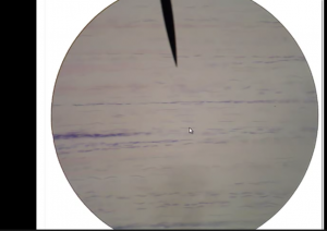
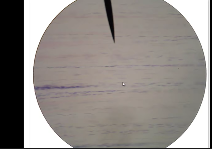
This is dense regular connective tissue. Notice the wood-like appearance, that’s a way to identify it. There is sub – microscopic collagen fibers which are all aligned and give this tissue it’s wood like appearance.
Collagen – a protein that provides structure to your bones, skin, tendons, and ligaments.
Fibroblasts- cells that are found in connective tissues.
We would find dense regular connective tissue in tendons and ligaments.
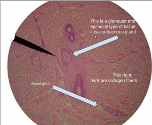
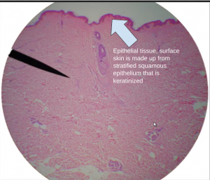
This is skin, the pinkish orange area contains collagen fibres, this time that aren’t running in the same direction, therefore it is dense irregular connective tissue. The big purple blotches are hair follicles, and are lined with epithelial tissue (what is the difference between epithelial and epithelium?) The white dot in the center of the hair follicle is the hair shaft. Same slide, smaller magnification.
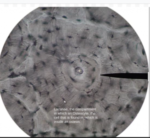
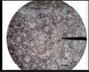
Bone tissue, the circles are called osteons which indicate that this is bone tissue.